
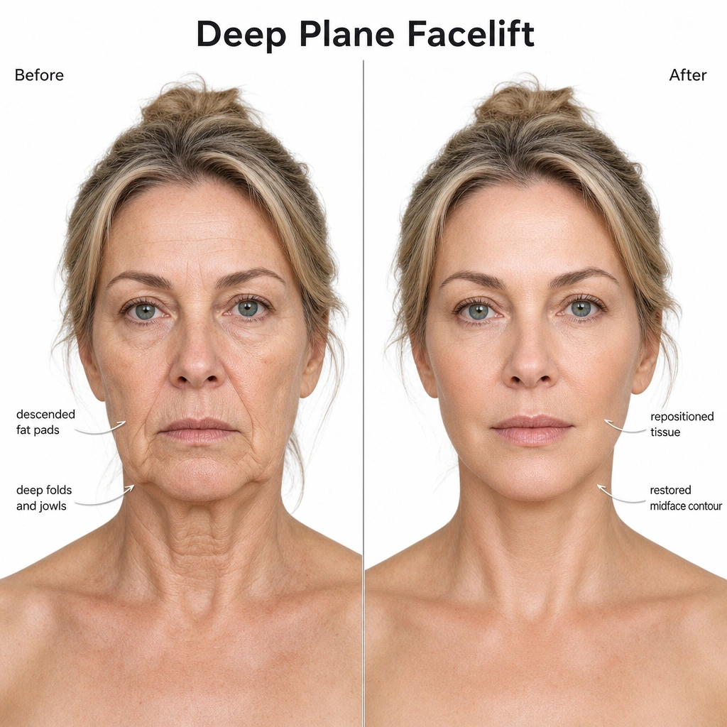
In one sentence: A deep plane facelift releases selected internal attachments and repositions the skin, fat, and SMAS support layer as one unit, so the correction is carried by deeper tissue rather than skin tension alone.
The deep plane facelift is defined by how tissue is dissected and moved, not simply by the fact that it addresses deeper facial layers than skin alone. This article sets out the surgical anatomy behind the technique — the composite flap concept, the retaining ligaments released, the facial nerve anatomy that governs dissection safety, and the sequence of steps involved — for readers who want the technical detail behind the procedure.
For candidacy, recovery timelines, and cost considerations, see the facelift procedure page.
What Is the Composite Flap in a Deep Plane Facelift?
In a standard subcutaneous facelift, the skin is dissected away from the SMAS (Superficial Musculo-Aponeurotic System) as a separate layer. The SMAS may then be tightened independently through plication or imbrication. A deep plane facelift instead dissects beneath the SMAS, elevating skin, subcutaneous fat, and SMAS together as a single composite flap. This anatomy-based approach was described in Hamra’s original paper on deep-plane rhytidectomy.
This distinction matters for two reasons:
- Vascular supply. The perforator vessels connecting the SMAS to the overlying skin remain intact within the composite flap, rather than being severed during a separate subcutaneous dissection.
- Mechanical unity. Because skin and SMAS move as one unit, the flap can be repositioned along the vector needed to correct descended tissue, rather than relying on skin tension to simulate a lift.
The composite flap concept is the anatomical basis for every other aspect of the technique discussed below.
Which Facial Retaining Ligaments Are Released, and Why?
Facial soft tissue is held in position by a series of fibrous retaining ligaments that anchor the dermis to the underlying periosteum or deep fascia. Their locations, connections, and variation have been documented in anatomical dissection studies of the facial retaining ligaments. A subcutaneous or SMAS-plication facelift largely works around these ligaments; a deep plane facelift releases selected ligaments directly, allowing the composite flap to move rather than merely stretch.
| Ligament | Anatomical location | Function | Relevance to deep plane release |
|---|---|---|---|
| Zygomatic cutaneous ligaments | Zygomatic arch / malar region | Anchor cheek skin to the zygoma | Release allows mid-face composite mobilization |
| Masseteric cutaneous ligaments | Anterior border of the masseter | Anchor lower cheek to the masseter fascia | Release allows jowl and lower-face repositioning |
| Mandibular (mandibulocutaneous) ligament | Anterior mandible | Anchor jawline skin to bone | Selectively addressed depending on jowl severity |
| Platysma-auricular ligament | Angle of mandible / neck | Anchors platysma to the deep neck fascia | Released when neck correction is combined |
Only the ligaments relevant to the individual’s pattern of descent are released — ligament release is targeted anatomical work, not a blanket dissection.
Layer-by-Layer Surgical Steps of Deep Plane Dissection
While specifics vary by anatomy and whether neck correction is combined, the deep plane technique generally follows this sequence:
- Skin incisions are marked along the temporal hairline, pre-auricular crease, and into the occipital scalp.
- A limited subcutaneous dissection is carried out only across the narrow anterior cheek margin, preserving SMAS attachment everywhere else.
- The SMAS is identified and entered, transitioning the dissection into the sub-SMAS (deep) plane.
- Sub-SMAS dissection proceeds anterior to the parotid gland, across the masseter.
- The zygomatic and masseteric cutaneous ligaments are released under direct visualization.
- The mobilized composite flap is repositioned along the vector required to correct the specific pattern of descent.
- The flap is fixed to stable points — typically the deep temporal fascia and periosteum — to carry the corrective tension internally.
- Skin is redraped over the repositioned composite flap and closed without tension, since the lift itself is already held by the internal fixation.
How Does Deep Plane Dissection Protect the Facial Nerve?
This is the anatomical question that governs why deep plane technique requires specific surgical training beyond standard facelift experience.
Over the body of the parotid gland, the facial nerve’s branches (frontal, zygomatic, buccal, and marginal mandibular) run deep to the SMAS. A dissection correctly maintained in the sub-SMAS plane over the parotid therefore stays superficial to these branches. As the branches exit the anterior border of the parotid and cross the masseter toward the mid-face, however, they become progressively more superficial — this region is treated as a defined anatomical danger zone. A 2023 cadaveric study of the deep fascia and facial nerve reinforces why dissection must remain at the most superficial level of the deep fascia.
Correct execution of the technique depends on:
- Precise identification of the sub-SMAS plane before crossing the anterior parotid border
- Recognition of the danger zone over the masseter, where dissection depth must be adjusted
- Direct visualization rather than blunt dissection through poorly defined tissue planes
This is why deep plane dissection is considered more technically demanding than subcutaneous or SMAS-plication facelifts, and why it is performed by surgeons with specific training in this anatomical plane. As with any surgical technique, correct execution reduces — but does not eliminate — inherent procedural risk.
Deep Plane vs. SMAS Plication vs. Mini Facelift
| Deep plane facelift | SMAS plication facelift | Mini facelift* | |
|---|---|---|---|
| What the surgeon does | Works beneath the SMAS and releases selected retaining ligaments; skin and SMAS move together | Folds and sutures the SMAS to tighten it, usually without broad deep-plane ligament release | Uses shorter incisions and a smaller area of dissection; the SMAS may still be plicated or otherwise treated |
| Commonly considered for | Moderate to significant cheek, jowl, and jawline descent; neck treatment may be added | Mild to moderate lower-face and jowl laxity, depending on anatomy and the surgeon’s technique | Early lower-face or jawline laxity when a limited correction is appropriate |
| Potential advantage | Greater composite-flap mobility can address the mid-face and lower face together | Effective structural support without the same extent of deep-plane dissection | Smaller operative field and often a shorter early recovery |
| Important limitation | More extensive, technically demanding dissection close to facial nerve anatomy | May provide less direct mid-face mobilization when retaining ligaments are not released | Limited correction; does not substitute for a full facelift when laxity is more advanced |
| Decision point | Best judged by the pattern of tissue descent and need for ligament release | Best judged by where support is needed and whether mid-face release is necessary | Best judged by whether the desired result can realistically be achieved with limited access |
*“Mini facelift” is not one standardized anatomical operation; it generally refers to a limited-incision, limited-dissection approach. A systematic review of limited-incision facelifts found multiple variations, which is why patients should ask exactly which layers and areas a proposed “mini” lift will treat.
Current comparative evidence does not show that one method is best for every patient. A 2025 systematic review and meta-analysis comparing SMAS and deep techniques found broadly comparable safety profiles, but limited direct comparisons and inconsistent outcome measures prevented firm conclusions about relative efficacy. The practical question is therefore not “Which name is best?” but “Which operation matches my anatomy, priorities, and acceptable recovery?” For more on candidacy and recovery, see the facelift procedure page.
Deep Plane Facelift Cost in Dubai
Facelift cost is determined by the actual surgical plan rather than a standard online figure. Relevant factors include whether correction is limited to the lower face or extends to the mid-face and neck, the duration and complexity of surgery, anaesthesia and facility requirements, previous facial surgery, pre-operative testing, and the follow-up included in the quotation.
“Lower face lift cost” and “mini face lift cost” are common search terms, but neither label describes one standardized operation. A lower-face or mini approach may involve a smaller operative field, yet cost cannot be compared responsibly until the surgeon states which tissue layers, facial regions, and associated neck procedures are included. Patients comparing facelift cost and recovery in Dubai should request an itemized quotation after examination rather than choose a technique from price alone.
Facelift Recovery Timeline After Deep Plane Rhytidectomy
Rhytidectomy recovery is individual, and the operating surgeon’s instructions take priority over any general timeline. The American Society of Plastic Surgeons’ facelift recovery guidance notes that dressings or drains may be used and that patients need individualized instructions for wound care, medication, follow-up, and return to activity.
- First week: swelling, bruising, tightness, and temporary altered sensation are expected. Early review focuses on the incisions, dressings or drains, and signs of bleeding or infection.
- Weeks two to four: visible bruising and swelling generally improve, but readiness for work or social events depends on the extent of surgery, the person’s job, and healing progress.
- Following months: residual edema, firmness, numbness, and incision color continue to settle. Research on lymphatic drainage after rhytidectomy supports that internal recovery can continue for months even after the face looks socially presentable.
This facelift recovery timeline is a planning framework, not a promise. Increasing pain or swelling, one-sided rapid enlargement, bleeding, fever, breathing difficulty, chest pain, or other unexpected symptoms require prompt contact with the surgical team or urgent medical assessment.
Facelift Scarring and Incision Care
Facelift scarring is unavoidable because the operation requires incisions. Their course depends on the surgical plan and may follow the temporal hairline, natural creases around the ear, the posterior hairline, and sometimes a short crease beneath the chin when neck treatment is included. Tension-free closure aims to keep the corrective load on deeper fixation rather than the skin.
Early incision care should follow the surgeon’s written instructions; patients should not start scar products, massage, lasers, or other treatments without approval. Scar color and firmness change gradually, and plastic-surgery scar maturation guidance describes maturation as a process that can take six months to a year. Smoking, infection, wound tension, sun exposure, individual scar biology, and previous surgery can affect the final appearance.
Complications Specific to Deep Plane Dissection
As with any surgical technique, deep plane facelift carries risks specific to its dissection plane and the tissue mobilized:
- Hematoma — bleeding beneath the composite flap, most often in the first 24–48 hours
- Temporary facial nerve neurapraxia — nerve stretch or bruising causing transient weakness, which typically resolves as swelling subsides
- Permanent nerve injury — rare, and associated with dissection outside the correct anatomical plane
- Contour irregularity — most often the result of incomplete ligament release rather than the composite flap technique itself
- Flap ischemia — uncommon given the preserved vascular supply of the composite flap, but a relevant consideration in smokers or previously scarred tissue
These risks, their likelihood, and mitigation are reviewed individually during a pre-operative clinical consultation; no surgical technique eliminates procedural risk entirely.
What Determines Candidacy for Composite Flap Elevation?
Composite flap elevation is not the appropriate technique for every patient. Candidacy depends on:
- The degree and pattern of mid-face and jawline descent
- Skin and soft tissue quality, including elasticity and thickness
- Any prior facial surgery affecting tissue planes or scarring
- General medical fitness for a longer operative and anaesthesia time relative to a subcutaneous or limited-incision lift
Patients with early or mild laxity are frequently better suited to a more limited technique; the anatomical assessment during consultation — not the desire for a specific named technique — determines what is appropriate.
Dr. Bouraoui Kotti, EBOPRAS-certified Consultant Plastic Surgeon at Seline Clinic, Jumeirah, Dubai, performs deep plane facelift surgery based on individual anatomical assessment. This article is provided for patient and professional education; it does not substitute for a clinical consultation.
How Should Deep Plane Facelift Before-and-After Images Be Assessed?
Deep plane facelift before-and-after photographs should be compared at similar angles, lighting, facial expression and follow-up intervals. Early swelling can obscure the final jawline and midface contour, so photographs from different healing stages should not be treated as equivalent. Review the clinical before-and-after gallery separately from the educational anatomy diagram on this page, which is not a patient result.
Medical disclaimer: This article is provided for educational purposes. Suitability for surgery, technique selection, and outcomes depend on individual anatomical factors, health status, and personalized surgical planning. A consultation with a DHA-licensed Consultant is required before any procedure.
Related procedures and guides:
- Facelift (Rhytidectomy) — Full procedure details, candidacy, and risks
- Facelift in Dubai — Candidacy, procedure details, and consultation information
- Facelift & Neck Contouring — Surgical procedure page
- Neck Lift (Cervicoplasty) — Candidacy, techniques, recovery, cost factors, and risks
- Eyelid Surgery (Blepharoplasty) — Periocular rejuvenation
- Before & After Gallery — Clinical results documentation
- Book a Consultation — Private clinical assessment at Seline Clinic, Jumeirah
Frequently Asked Questions
What is a composite flap in a deep plane facelift?
A composite flap is the skin, subcutaneous fat, and SMAS elevated together as a single anatomical unit, rather than dissecting the skin and SMAS as separate layers. Because the natural attachments between these layers are preserved, the flap retains its own blood supply and can be repositioned as one structure along its original vector.
Which facial ligaments are released during deep plane facelift surgery?
The zygomatic cutaneous and masseteric cutaneous ligaments are released to mobilize the mid-face composite. When neck correction is combined with the facelift, the platysma-auricular ligament is also addressed. Releasing these retaining ligaments, rather than only tensioning the skin, is what allows descended tissue to be repositioned rather than simply pulled tighter.
How does deep plane dissection protect the facial nerve?
Over the parotid gland, the facial nerve branches run deep to the SMAS, so a correctly maintained sub-SMAS plane stays superficial to them. The branches become more superficial as they cross the masseter toward the mid-face, which is why this region is treated as a defined anatomical danger zone requiring precise plane identification and surgical experience with the technique.
Why is the deep plane technique more vascularly stable than a subcutaneous facelift?
A subcutaneous flap depends on a random subdermal blood supply, which thins as the flap is elevated. A composite flap keeps its myocutaneous perforator vessels intact because the SMAS is elevated together with the skin, supporting flap perfusion — a relevant consideration in smokers or previously operated tissue.
What determines candidacy for composite flap elevation?
Candidacy depends on the degree of mid-face and jawline descent, skin and soft tissue quality, prior facial surgery, and general medical fitness for a longer operative and anaesthesia time compared with a subcutaneous or limited-incision lift. Anatomical assessment during a clinical consultation determines whether composite flap elevation is appropriate.
What are the technique-specific risks of deep plane facelift surgery?
Documented risks include hematoma, temporary facial nerve neurapraxia, and, rarely, permanent nerve injury if the dissection plane is not correctly maintained. Incomplete ligament release can also result in residual contour irregularity. These risks are inherent to the technique and are discussed individually during pre-operative consultation.
How is the composite flap secured during deep plane facelift surgery?
The mobilized composite flap is fixed to stable points such as the deep temporal fascia and periosteum along a vector determined by the direction of tissue descent, then the skin is redraped and closed without tension. Because the lift is carried by the fixed composite flap rather than skin tension, the closure itself does not bear the corrective load.
How much does a deep plane facelift cost in Dubai?
There is no universal price. Cost depends on the extent of facial and neck correction, operating and anaesthesia requirements, facility fees, surgeon expertise, pre-operative assessment, and follow-up care. A clinical examination is required before an itemized quotation can be provided.
What is the typical facelift recovery timeline?
Recovery varies with the extent of surgery and individual healing. Bruising, swelling, tightness, and temporary numbness are expected early; many patients plan roughly two to four weeks away from major social commitments, while residual swelling, sensation, and scars continue to mature over several months. The operating surgeon's instructions take priority.
Where are deep plane facelift scars placed?
Incisions are generally planned around the temporal hairline, natural contours around the ear, and sometimes the posterior hairline or beneath the chin when neck work is included. Scars are permanent but usually fade as they mature; their position and appearance depend on the operative plan and individual healing.
Popular Procedures
Specialties
Related Clinical Articles
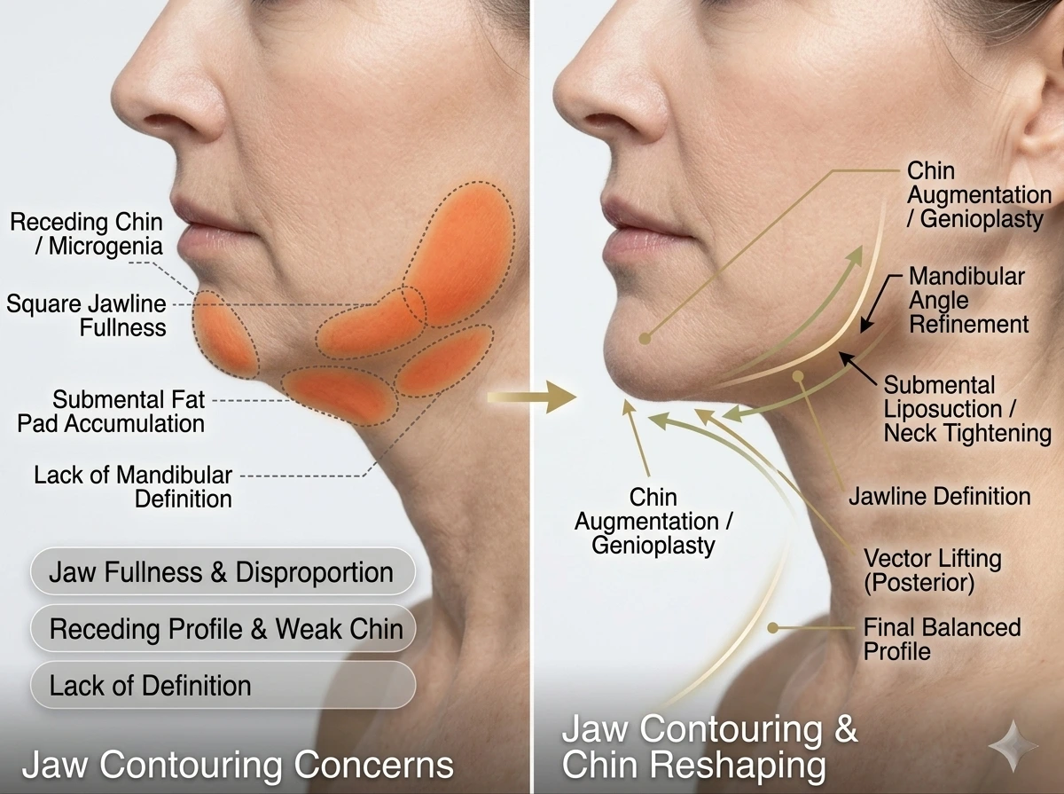
What's the Difference? Jaw Contouring and Jaw Reduction
Jaw contouring may reshape bone or soft tissue, while jaw reduction surgically narrows selected bone. Compare options, candidacy, recovery, risks and cost factors.
Aug 18, 2026
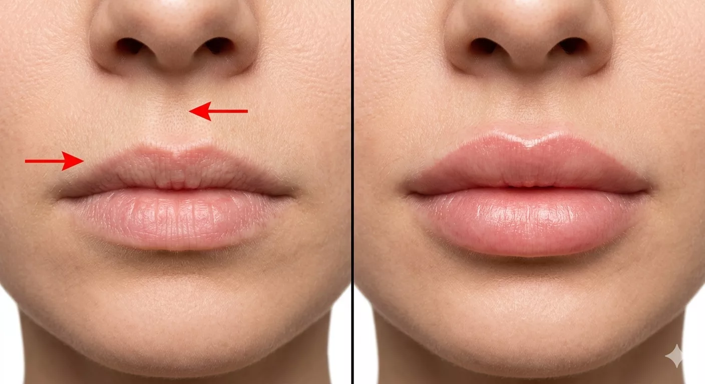
Lip Lift Surgery in Dubai: Scars, Recovery and Options
Considering lip lift surgery in Dubai? Learn about candidacy, scars and recovery, then compare alternatives. Dr. Kotti discusses options in consultation today.
Jun 28, 2026
